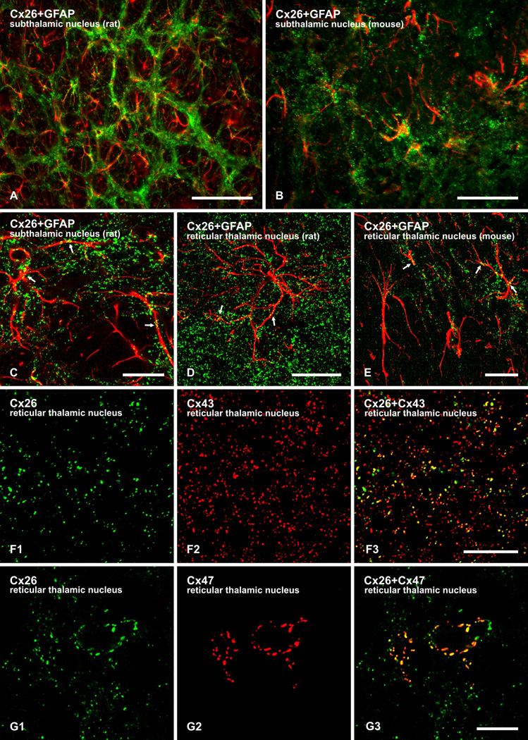
FIG. 4.
Immunofluorescence localization of labelling for Cx26 with GFAP, Cx43 and Cx47 in brain. (A,B) Low magnification overlays showing co-distribution of Cx26 (green) with GFAP-positive astrocytes (red) in rat (A) and mouse (B) brain. (C–E) Higher magnification confocal overlay images showing co-localization of punctate labelling for Cx26 (green) along GFAP-positive astrocyte processes (red) in the subthalamic nucleus (C) and reticular thalamic nucleus (D) of rat, and the reticular thalamic nucleus of mouse (E), with red/green overlap seen as yellow (arrows). (F) Laser scanning confocal double immunofluorescence labelling for Cx26 with Cx43 in mouse reticular thalamic nucleus, showing Cx26-positive puncta (F1) co-localized with Cx43-positive puncta (F2), as seen by yellow in overlays (F3). Laser scanning confocal double immunofluorescence labelling for Cx26 with Cx47 in mouse reticular thalamic nucleus, showing Cx26-positive puncta (G1) co-localized with Cx47-positive puncta (G2), as seen by yellow in overlays (G3). Scale bars: A, 100 µm; B, 50 µm; C–E, F,G, 10 µm.

