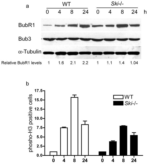
Figure 6. Spindle Checkpoint activation in WT and Ski−/− MEFs.
WT and Ski−/− MEFs were treated with nocodazole (125ng/ml) for 0 to 24 hours. Levels of BubR1 and Bub3 were assessed by western blotting (a). BubR1 levels are expressed as relative to time 0h (untreated cells). A representative experiment is shown. (b) Cells positive for phosho-H3(S10) were determined by flow cytometry. Values are expressed as fold increase relative to time 0h (untreated cells).

