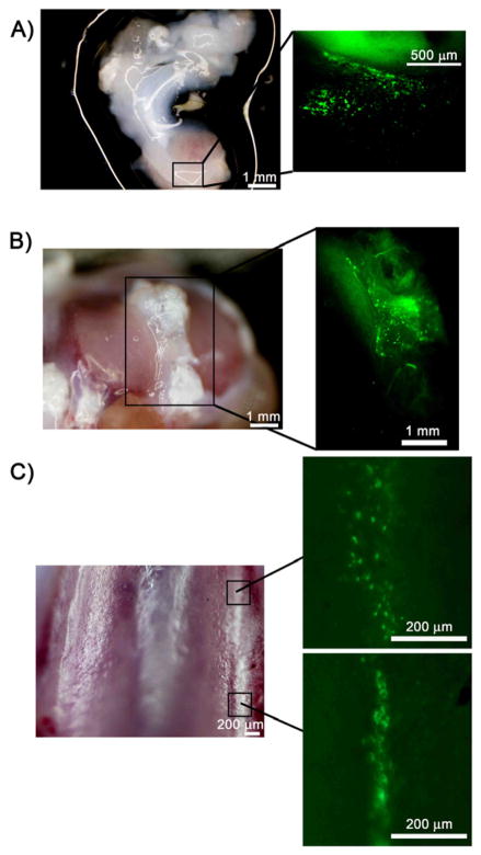
Figure 4.
GFP+ cells in tissues harvested from the right knee joint of rats injected with AAV-CMV-GFP and imaged with a stereomicroscope. (A) Brightfield image (left panel) of meniscal tissue with the area located within the square enlarged to show the GFP+ cells (right panel). (B) Brightfield image (left panel) of tibial plateau. Area within the square is magnified in the right panel and shows GFP+ cells within the soft tissue. (C) Brightfield image of the trochlea (left panel). GFP+ cells were sparse in articular cartilage. Two small areas with GFP+ cells were found on the trochlear ridge in this animal (right panels).

