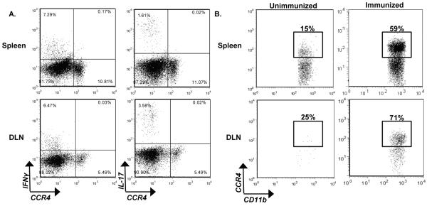
Figure 6. CCR4 is expressed by iMϕ post immunization.
WT mice were immunized and killed at peak clinical disease. CCR4 expression on pathogenic cell populations was evaluated by flow cytometric analysis. (A) Representative flow dot plots of WT spleens and DLN displaying IFNγ and IL-17 versus CCR4. The data is derived from a combined forward versus side-scatter and CD45+CD4+ gate. (B) Representative flow dot plots from unimmunized and immunized WT mice, illustrate CCR4 expression by CD11b+Ly6Chi cells in the spleen and DLN. The data is derived from a CD45+CD11b+Ly6Chi gate. The data is representative of two experiments, n=3 mice/group.

