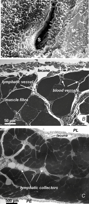
Figure 1. Arrangement of the diaphragmatic lymphatic network.

A, scanning electron photomicrograph of an elliptical lymphatic stoma on the tendinous pleural surface of the rat diaphragm. The stoma (S), delimited by a net border, opens at the confluence between adjacent mesothelial cells (MC) characterized by a mesh of microvilli protruding from the cell surface. B, semi-thin cross-section of rat pleural hemi-diaphragm stained with crystal violet and basic fuchsin showing the arrangement of the diaphragmatic lymphatic network originating from the pleural mesothelial surface. Lymphatic submesothelial lacunae located within the interstitial space beneath the mesothelial layer are in continuity with transverse lymphatic vessels (arrow) located in the deep interstitial space and running perpendicularly to the lacunae and through the diaphragmatic skeletal muscular fibres. C, semi-thin cross-section of rat entire diaphragm stained with crystal violet and basic fuchsin, showing both the pleural (PL) and the peritoneal (PE) mesothelial surfaces. Transverse lymphatic vessels departing from the pleural and peritoneal submesothelial lacunae empty into central lymphatic collectors, located in the deep interstitial space, which receive the lymph collected from both surfaces of the diaphragm.
