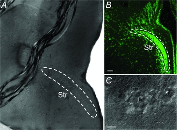
Figure 1. Anatomical location of the striatum (Str).

A, coronal acute brain slice in the recording chamber showing the striatal cell band inside the white lines. B, fluorescent Nissl staining of a coronal section displaying the band of striatal cell bodies. C, photograph of individual striatal neurons in the recording chamber. Scale bars = 100 μm in A and B, 15 μm in C.
