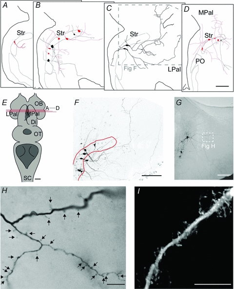
Figure 8. Morphology and topography of striatal neurons.

A–D, schematic representations of IRNs (pale) and non-IRNs (black), in sections from rostral to caudal according to E. E, overview of the lamprey brain, including the level of coronal sectioning shown in A–D. F, confocal image showing the morphological properties of the two non-IRNs shown in C, among 4 other labelled but unrecorded neurons. See inset in C for the location in the coronal section. G, overview of a spiny IRN by light microscopy of a DAB-stained slice. H, close-up of spiny dendrites in the IRN shown in G, according to white inset. I, confocal image of another spiny dendrite labelled in the striatum. Scale bars = 100 μm in A–D, F and G; 1 mm in E; 10 μm in H and I. Abbreviations are as follows: Di, diencephalon; LPal, lateral pallium; MPal, medial pallium; OB, olfactory bulb; PO, nucleus preopticus; OT, optic tectum; Str, striatum.
