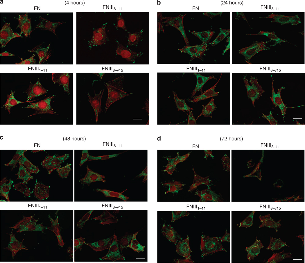
Figure 4. Fibronectin (FN)-null cells spread and formed focal contacts on FN-central cell-binding domain (FNIII8–11) similar to cells on intact FN or contiguous arrays of FN-CCBD and FN-growth factor-binding domains (GFBDs) (FNIII1–11 or FNIII8–v15) for at least 24 hours.
FN-null fibroblasts were cultured at 8,000 cells per 35-mm dish that had been precoated with 0.125 µm intact FN or glutathione S-transferase (GST)-tagged FNIII8–11, FNIII1–11, or FNIII8–v15 in DMEM for 4, 24, 48, or 72 hours at 37 °C. After the appropriate incubation times, cells were fixed, permeabilized, and stained for vinculin and actin as described in Materials and Methods. (a) FN-null cells had formed vinculin-positive focal contacts (green) and actin stress fibers (red) on all precoated surfaces by 4 hours. (b) Vinculin-positive focal contacts and actin stress fibers persisted in all cells at 24 hours. (c) By 48 hours, FN-null cells plated on GST-FNIII8–11 were less well spread and had only a few vinculin-positive focal contacts, whereas cells on intact FN, FNIII1–11, or FNIII8–v15 remained well spread with prominent actin stress fibers and many focal contacts. (d) By 72 hours, FN-null cells on dishes coated with GST-FNIII8–11 had become sparse and those that remained were mostly rounded with minimal evidence of actin bundles or focal contacts, whereas cells on intact FN, FNIII1–11, or FNIII8–v15 continued to be spread with prominent actin stress fibers and focal contacts. Bar = 20 µm.

