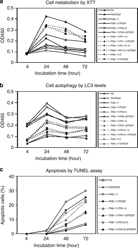
Figure 6. Surface-bound fibronectin (FN)-growth factor-binding domains (GFBDs) in noncontiguous arrays with the FN-central cell-binding domain (FNIII8–11) supported FN-null fibroblast responsiveness to platelet-derived growth factor-BB (PDGF-BB).
FN-null fibroblasts were cultured at 4,000 cells per well in 96-well plates precoated with 0.125 µm intact FN or glutathione S-transferase (GST)-tagged FN domains in DMEM for 4 hours, and then incubated with DMEM and 1% BSA with or without 30 ng ml−1 PDGF-BB at 37 °C for times indicated. (a) Survival as judged by the XTT assay. (b) Autophagy as judged by ELISA for light chain 3 (LC3). (c) Apoptosis as determined by the TUNEL assay. Percent apoptosis was determined by counting those cells positive for DNA fragmentation, dividing by total cell number, and multiplying by 100. Fifty cells were counted in three replicate plates. Data points indicate mean ± SD. The data shown in each of these panels are representative of at least three different experiments. All recombinant FN domains were GST-tagged but the prefix is omitted here for brevity. OD, optical density.

