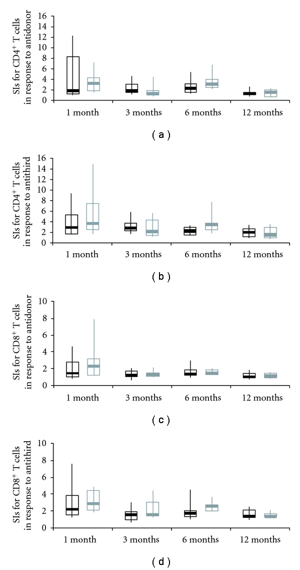
Figure 3.

Kinetics of stimulation index in the RI group and non-RI group during the first year after transplantation. Stimulation index (SI) of each of the CD4+ T-cell (a, b) and CD8+ T-cell (c, d) subsets in the antidonor (a, c) and anti-third-party (b, d) MLR in patients in non-RI group (black line) and RI group (gray line). CD4+ and CD8+ T-cell proliferation and their SIs were quantified as follows. The number of division precursors was extrapolated from the number of daughter cells of each division, and the number of mitotic events in each of the CD4+ and CD8+ T-cell subsets was calculated. Using these values, the mitotic index was calculated by dividing the total number of mitotic events by the total number of precursors. The SIs of allogeneic combinations were calculated by dividing the mitotic index of a particular allogeneic combination by that of the self-control. The box plot represents the 25th to 75th percentile, the dark line is the median, and the extended bars represent the 10th to the 90th percentile.
