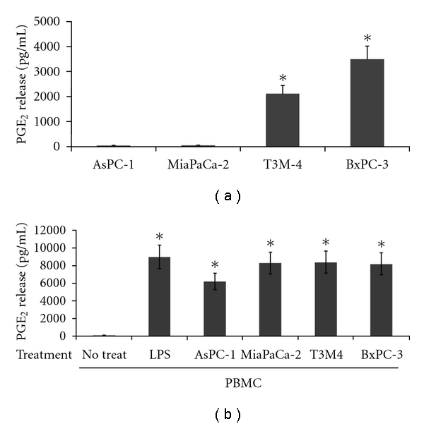
Figure 1.

Release of PGE2 from pancreatic carcinoma cells and peripheral blood mononuclear cells. (a) PGE2 production from PDAC monocultures of cells. Cells (AsPC-1, MiaPaCa-2, T3M-4, BxPC-3) were plated in 48-well plates at a density of 2.5 × 10⁴ cells/well in 500 μL of medium/well, and supernatants were collected at 48 hrs for PGE2 ELISA measurement. (b) PGE2 production from PBMCs. After purification over a Histopaque gradient mononuclear cells were plated in a 48-well plate (5 × 10⁵ cells/well) either alone or onto preseeded PDAC cultures (2.5 × 10⁴ cells/well). 48 hours after coculturing the cells, supernatants were collected and analysed for PGE2 expression. PBMCs treated with LPS and ConA served as controls. All experiments were repeated in triplicate with three different healthy blood donors. *Standard deviations were calculated, and results were considered significant with P values from Student's t-test below 0.05.
