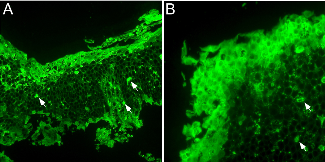
Figure 1.
Deposition of EDN in esophageal tissue from patients with eosinophilic esophagitis (EoE). Esophageal biopsy specimens from two separate EoE patients (A and B) were stained with rabbit anti-human EDN antibody followed by FITC-conjugated goat anti-rabbit IgG. Wide spread diffuse extracellular EDN is observed throughout the esophageal epithelium, in particular on the luminal surface and in the superficial mucosa. Arrows indicate intact eosinophils. Original magnification; ×160 (A) and ×400 (B).

