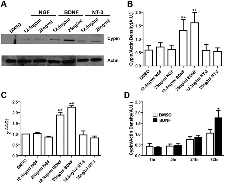
Figure 1.
Endogenous cypin expression increases in response to BDNF but not NGF or NT-3. A, Cells were treated with the indicated concentrations of neurotrophins beginning at 7 DIV for 72 h. Extracts from untreated and treated cultures of hippocampal neurons were analyzed by SDS-PAGE and Western blotting using antibodies that recognize cypin or actin. Representative blot is shown. B, Densitometry analysis of cypin normalized to actin expression. C, Quantitative RT-PCR assay for the rat cypin gene (GDA) after indicated treatments of cultured hippocampal neurons. **p < 0.01, determined by ANOVA followed by Student-Newman–Keuls multiple-comparison test compared to DMSO as control. Cypin mRNA and protein levels are upregulated by increasing doses of BDNF. D, Exposure of hippocampal neurons to BDNF for 72 h, but not 24 h or less, results in increased cypin levels. Cells were treated with BDNF at 7 DIV for each indicated duration. Cypin protein levels were compared by Western blotting. *p < 0.05 by two-tailed t test compared to control. Error bars indicate SEM. A.U., Arbitrary units; n = 3 experiments for all panels.

