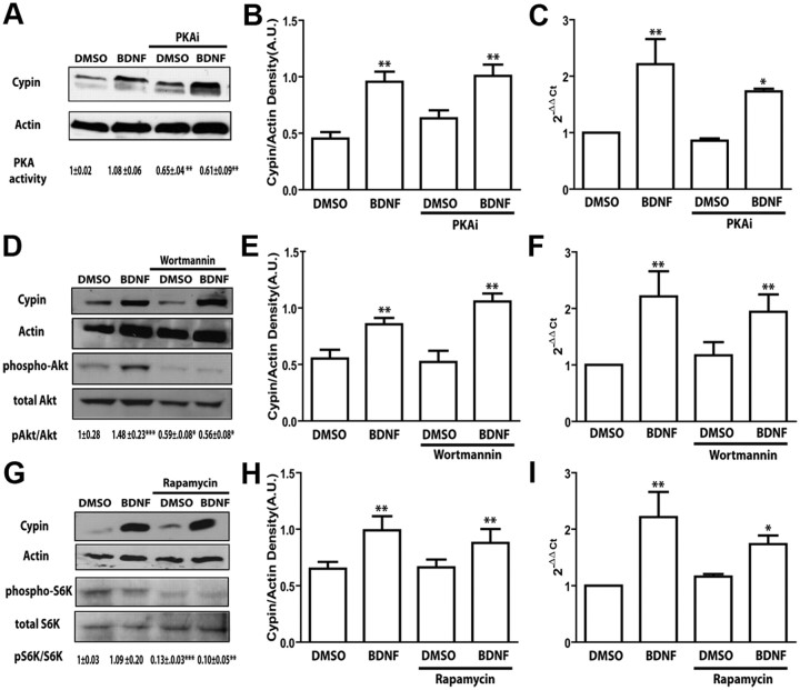
Figure 3.
The cAMP/PKA, PI3K/Akt/PKB, and mTOR pathways are not involved in BDNF-mediated increases in cypin expression. A–I, Hippocampal neurons were treated with membrane-permeable PKA inhibitory (PKAi) peptide (25 ng/ml; Myr-N-Gly-Arg-Thr-Gly-Arg-Arg-Asn-Ala-Ile-NH2) (A–C), wortmannin (100 nm) (D–F), or rapamycin (25 ng/ml) (G–I) at 7 DIV and treated with BDNF for 72 h. Cytoplasmic protein extracts were resolved by SDS-PAGE and Western blotting using antibodies that recognize indicative proteins. PKA activity was measured by absorbance at 450 nm with an ELISA-based PKA activity assay. PKA activity is shown as mean normalized absorbance ± SEM. Densitometric analysis of active phospho-proteins normalized to total proteins are indicated as mean ± SEM. Densitometric analysis of cypin is normalized to actin protein expression. Extracts from treated cultures were then analyzed by quantitative RT-PCR using specific rat cypin/GDA primers. *p < 0.05, **p < 0.01, ***p < 0.001 by ANOVA followed Student-Newman–Keuls multiple-comparison test compared to DMSO as vehicle control. Error bars indicate SEM. n = 3 experiments for all panels with representative blots shown in A, D, G. S6K, p70S6 kinase.

