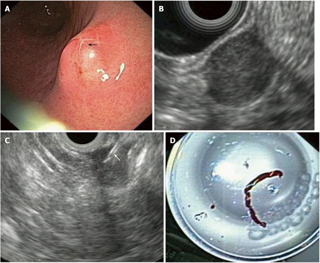
Figure 2.

Gastric gastrointestinal stromal cell tumors: Endoscopic aspects, endoscopic ultrasonography-imaging and tissue sampling. A: Endoscopic image of the lesion; note that the lesion is covered by a normal mucosa with a central umbilication (black arrow); B: EUS imaging of the lesion, which is located in the 4th echo-poor layer (muscularis propria); C: EUS-FNA of the lesion; note the presence of the needle (white arrow); D: Histological specimen of the EUS-FNA. EUS:Endoscopic ultrasonography; EUS-FNA: EUS-guided fne needle aspiration.
