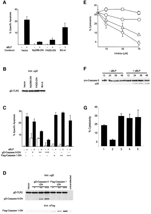
Fig. 4. TLR2-induced apoptosis proceeds through a FADD/caspase 8 pathway. (A) TLR2-mediated apoptosis is inhibited by FADD-DN but not by Bcl-xl. 293 cells were transiently co-transfected with 0.25 µg of an expression plasmid encoding gD-TLR2 and 0.3 µg of expression plasmids encoding the indicated dominant negatives or Bcl-xl. At 24 h post-transfection, the cells were incubated with or without 1 µg/ml sBLP as indicated and apoptosis was determined by TUNEL staining. (B) Anti-gD western blot of lysates prepared from cells transfected as in (A) indicating equivalent expression of gD-TLR2 among transfections. (C) TLR2-mediated apoptosis is inhibited by a catalytically inactive mutant of caspase 8. 293 cells were transiently co-transfected with 0.15 µg of an expression plasmid encoding gD-TLR2 and either 0.05 (+), 0.15 (++) or 0.45 µg (+++) of expression plasmids encoding gD-caspase 8-DN or Flag-caspase 1-DN. Apoptosis was induced and quantified as in (A). Data in (A) and (C) are reported as the percentage specific apoptosis relative to cells transfected with gD-TLR2 without sBLP. (D) Anti-gD (upper panel) and anti-Flag (lower panel) western blots of lysates prepared from cells transfected as in (C). (E) sBLP-mediated cell death of THP-1 cells is attenuated by a peptide inhibitor of caspase 8. THP-1 cells were pre-incubated with medium containing 75 µg/ml cycloheximide without (star) or with the indicated concentrations of z-FA-fmk (squares), Ac-YVAD-cmk (circles), z-IETD-fmk (triangles) or z-VAD-fmk (diamonds). sBLP was added to a final concentration of 2 ng/ml. Some standard deviations are within the limits of the data points. (F) Anti-casapse-8 western blots of lysates prepared from 293 cells co-transfected with gD-TLR2 and NIK-DN and incubated for the indicated times (h) with sBLP (1 µg/ml). Pro-caspase 8 (p55) and the p28 proteolytic intermediate are shown. (G) sBLP-mediated cell death is not mediated through TNF or Fas. THP-1 cells were pre-incubated for 4.5 h with medium only (1) or with 5 µg/ml anti-TLR2 mAb 2392 (2), an isotype-matched control mAb (3), anti-Fas (4) or anti-TNF mAb (5) and then with 100 pg/ml sBLP. Cytotoxicity was assayed by LDH release.

An official website of the United States government
Here's how you know
Official websites use .gov
A
.gov website belongs to an official
government organization in the United States.
Secure .gov websites use HTTPS
A lock (
) or https:// means you've safely
connected to the .gov website. Share sensitive
information only on official, secure websites.
