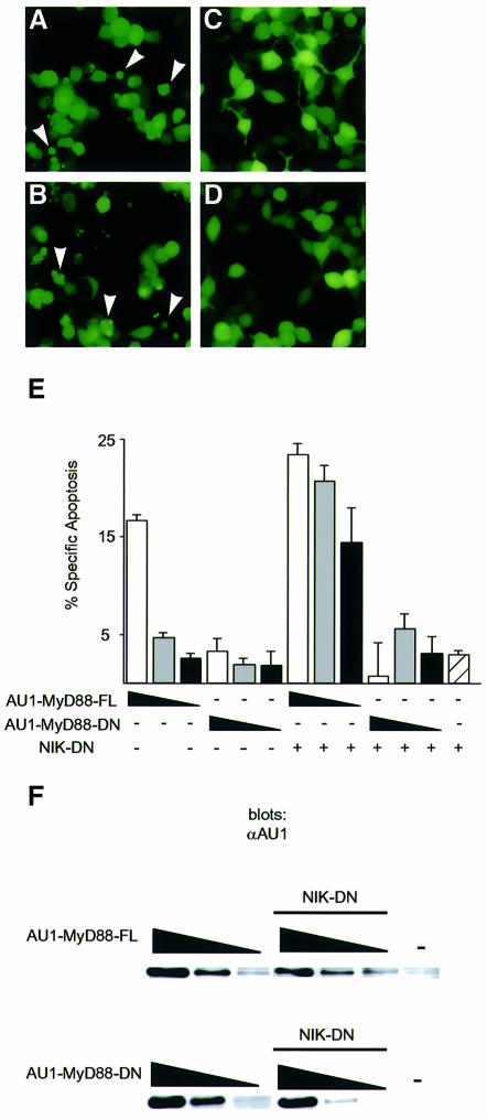
Fig. 5. MyD88-FL, but not MyD88-DN, induces apoptosis in 293 cells. (A–D) 293 cells were transiently transfected with 0.25 µg of expression plasmids encoding AU1-MyD88-FL (A and B) or AU1-MyD88-DN (C and D). In (B) and (D), 0.25 µg of an expression plasmid encoding NIK-DN was co-transfected. A plasmid encoding GFP (0.025 µg) was included in the transfection to label transfected cells. GFP-positive cells were photographed 48 h post-transfection. Arrowheads indicate cells with apoptotic morphology. (E) Inhibition of the NF-κB pathway facilitates MyD88-mediated apoptosis. 293 cells were transiently transfected with 0.25 (white bars), 0.1 (gray), 0.04 (black) or 0 µg (diagonal lines) of expression plasmids encoding AU1-MyD88-FL or AU1-MyD88-DN. Where indicated, 0.25 µg of an expression plasmid encoding NIK-DN was co-transfected. A plasmid encoding GFP (0.025 µg) was included to label transfected cells. At 60 h post-transfection, apoptosis was determined by annexin V staining. The data are reported as the percentage specific apoptosis in the GFP-positive (transfected) population relative to cells transfected with vector DNA. A specific increase in apoptosis was only detected in the GFP-positive population. (F) Anti-AU1 western blot of lysates prepared from cells transfected as in (E).

An official website of the United States government
Here's how you know
Official websites use .gov
A
.gov website belongs to an official
government organization in the United States.
Secure .gov websites use HTTPS
A lock (
) or https:// means you've safely
connected to the .gov website. Share sensitive
information only on official, secure websites.
