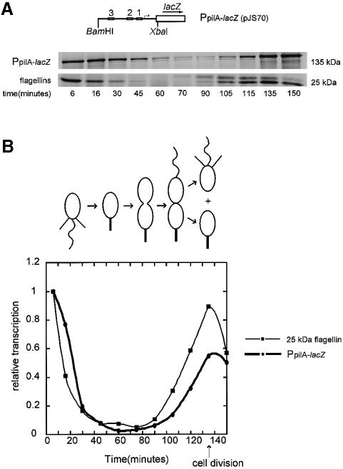
Fig. 8. Transcription of pilA is cell cycle controlled. (A) Diagram of the PpilA–lacZ fusion construct, pJS70. Wild-type C.crescentus strain NA1000 carrying pJS70 was synchronized and allowed to proceed through the cell cycle. The synthesis of β-galactosidase was monitored by pulse labeling cells with [35S]methionine for 5 min at the times indicated and immunoprecipitating with anti-β-galactosidase antibody. Labeled β-galactosidase was resolved on a 10% SDS–PAGE gel. As a control, the flagellin subunits were immunoprecipitated from the same labeled cell extracts. (B) The β-galactosidase and the 25 kDa flagellin bands were quantitated using a phosphoimager and were normalized to the value of the most intense band in each series. A diagram of the Caulobacter cell cycle is shown. The pattern of pilA transcription coincides with the pattern of 25 kDa flagellin gene transcription and is similar to the timing of pilus assembly observed by electron microscopy.

An official website of the United States government
Here's how you know
Official websites use .gov
A
.gov website belongs to an official
government organization in the United States.
Secure .gov websites use HTTPS
A lock (
) or https:// means you've safely
connected to the .gov website. Share sensitive
information only on official, secure websites.
