
Figure 1.
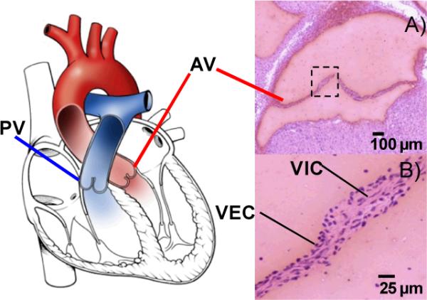
Illustration of the pulmonary (PV) and aortic valves (AV) within the heart. A) Hematoxylin and eosin stain of a coronal section from adult mouse aortic valve; B) Cellular anatomy of the valve. VEC-valvular endothelial cells, VIC- valvular interstitial cells.