Figure 1.

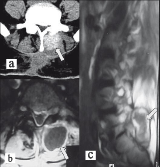
Lumbar imaging of our case revealed a paravertebral mass lesion located in the left side of the previous operation site (arrows). (a) Axial CT scan, (b) axial post-contrast enhanced MRI, (c) sagittal T2-weighted MRI

Lumbar imaging of our case revealed a paravertebral mass lesion located in the left side of the previous operation site (arrows). (a) Axial CT scan, (b) axial post-contrast enhanced MRI, (c) sagittal T2-weighted MRI