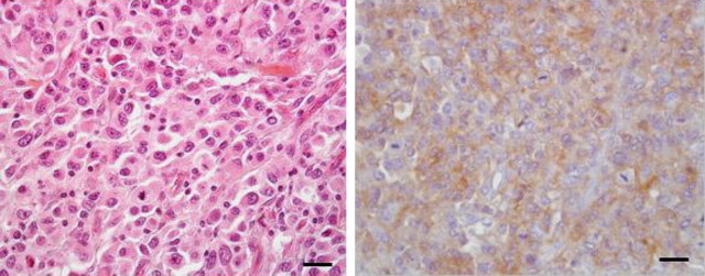
Figure 2.
Histopathological and immunohistochemical features of HS. Left: neoplastic proliferation of a pleomorphic population of large round uni- or multinucleated cells, with marked atypia and high mitotic index; hemalun–eosin staining; bar = 30 μm. Right: neoplastic cells expressing the leukocyte CD18 marker (immunoperoxidase reaction, diaminobenzidine as chromogen, hematoxylin counterstaining); bar = 30 μm. (This figure appears in color in the online version of Journal of Heredity.)

