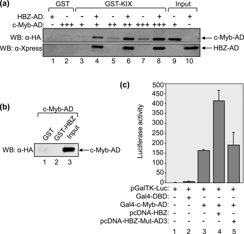
Figure 6. HBZ-AD enhances the binding of c-Myb-AD to KIX.
(a) HBZ-AD enhances the binding of c-Myb-AD to KIX in GST pull-down assays. Binding reactions contained GST-KIX (250 nM, lanes 3-8) and increasing concentrations of c-Myb-AD (0.1 μM, lanes 3 and 4; 0.25 μM, lanes 5 and 6; 0.5 μM; lanes 7 and 8). Lanes 4, 6 and 8 contained 7.5 μM HBZ-AD. Control reactions contained GST (250 nM) with HBZ-AD (7.5 μM, lane 1) or c-Myb-AD (0.5 μM, lane 2) as indicated. Fractions of c-Myb-AD (1%) and HBZ-AD (0.8%) inputs are shown in lanes 9 and 10, respectively. Bound proteins were resolved by SDS-PAGE and analyzed by Western blot (WB) using the antibodies indicated. (b) c-Myb-AD does not bind GST-HBZ in GST pull-down assays. Binding reactions contained c-Myb-AD (0.5 μM) and GST (7.5 μM, lane 1) or GST-HBZ (7.5 μM, lane 2). A fraction of the input of c-Myb-AD (1%) is shown in lane 3. (c) HBZAD increases transcriptional activation by c-Myb-AD in cells. Jurkat cells were transfected with pGalTK-Luc (100 ng) alone or in combination with Gal4-DBD (50 ng), Gal4-c-Myb-AD (50 ng), pcDNA-HBZ (100 ng) or pcDNA-HBZ-Mut-AD3 (100 ng) as indicated. Luciferase assays were performed 24 hours after transfection. The reported values are the average luminescence ± S.E. from one experiment performed in duplicate and are representative of three independent experiments.

