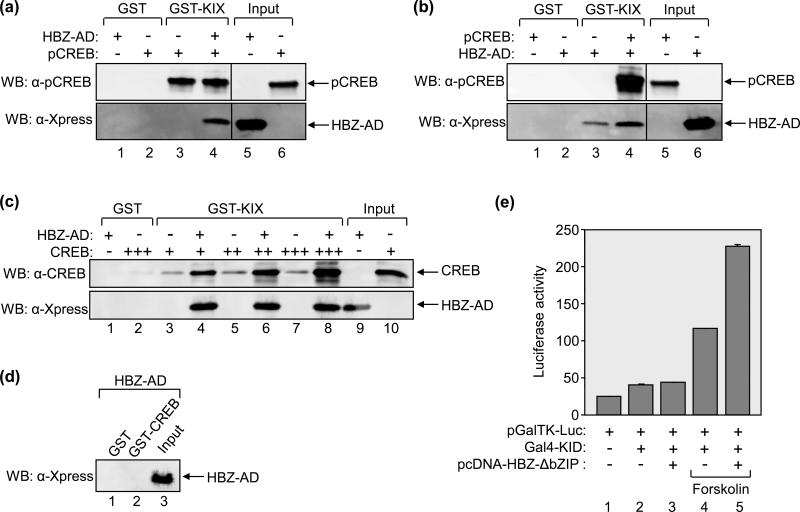
Figure 7. HBZ-AD affects the binding of CREB to KIX.
(a) HBZ-AD does not enhance pCREB-binding to KIX in GST pull-down assays. Binding reactions contained GST-KIX (25 nM, lanes 3 and 4) and pCREB (25 nM, lanes 3 and 4) in the absence or presence of HBZ-AD (1 μM, lane 4). Control reactions contained GST (25 nM) with HBZ-AD (1 μM, lane 1) or pCREB (25 nM, lane 2). Fractions of HBZ-AD (3.3%) and pCREB (24%) inputs are shown in lanes 5 and 6, respectively. Lanes shown are from the same membrane. Vertical lines show where the membrane was cropped. (b) pCREB enhances HBZ-AD-binding to KIX in GST pull-down assays. Binding reactions contained GST-KIX (25 nM, lanes 3 and 4) and HBZ-AD (25 nM, lanes 3 and 4) in the absence or presence of pCREB (100 nM, lane 4). Control reactions contained GST (25 nM) with pCREB (100 nM, lane 1) or HBZ-AD (25 nM, lane 2). Inputs of pCREB (6%) and HBZ-AD (130%) are shown in lanes 5 and 6, respectively. Lanes shown are from the same membrane. Vertical lines show where the membrane was cropped. (c) HBZ-AD enhances the binding of unphosphorylated CREB to KIX in vitro. Binding reactions contained GST-KIX (500 nM, lanes 3-8) and increasing concentrations of CREB (50 nM, lanes 3 and 4; 100 nM, lanes 5 and 6; 200 nM CREB, lanes 7 and 8). Lanes 4, 6 and 8 contained 5 μM HBZ-AD. Control reactions contained GST (500 nM) with HBZ-AD (5 μM, lane 1) or CREB (200 nM, lane 2). A fraction of HBZ-AD (2%) and CREB (12%) inputs are shown in lanes 9 and 10, respectively. (d) CREB does not bind HBZ-AD in GST pull-down assays. Binding reactions contained 5 μM HBZ-AD with either GST (200 nM, lane 1) or GST-CREB (200 nM, lane 2). A fraction of the input of HBZ-AD (2%) is shown in lane 3. (e) HBZ-AD increases transcriptional activation by pKID in cells. 293T/17 cells were transfected with pGalTK-Luc (100 ng) alone or in combination with Gal4-KID (100 ng), pcDNA-HBZ-ΔbZIP (100 ng) as indicated. Cells were treated with forskolin (2 μM) for 5h where indicated before the assays were performed. Luciferase assays were performed 24 hours after transfection. The reported values are the average luminescence ± S.E. from one experiment performed in duplicate and are representative of three independent experiments.

