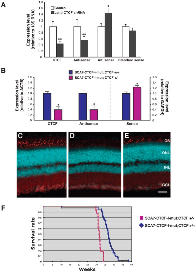
Figure 5. CTCF regulates promoter usage for ataxin-7 sense and antisense transcripts.
A) CTCF knock-down in Y-79 retinoblastoma cells leads to decreased ataxin-7 antisense RNA expression and increased exon 2A transcript expression. Human Y-79 retinoblastoma cells were infected with a lentiviral expression construct containing two tandem shRNAs targeting CTCF (lenti-CTCF-shRNA) or with empty vector (Control), and expression levels were measured by qPCR. Marked reductions in expression of CTCF and ataxin-7 antisense SCAANT1 transcripts were noted (p < 0.01 by ANOVA), and were accompanied by a significant increase in the ataxin-7 alternative sense transcript (p < .0.05 by ANOVA). Knock-down of CTCF did not affect expression of the ataxin-7 transcript initiated from a human-specific ataxin-7 promoter located >40 kb 5′ to the alternative promoter. Experiments were performed in triplicate, the expression level for each transcript upon CTCF knock-down is normalized to its respective control, and error bars = s.e.m.
B) Decreased CTCF dosage reduces SCAANT1 expression in SCA7-CTCF-I-mut mice. We crossed SCA7-CTCF-I-mut mice with CTCF +/− mice and derived SCA7-CTCF-I-mut mice on a CTCF heterozygous null background (CTCF +/−) or a wild-type background (CTCF +/+). We performed qRT-PCR analysis of CTCF in the brain of three month-old mice, and confirmed reduced expression of CTCF in SCA7-CTCF-I-mut; CTCF+/− mice (p < .0.05 by ANOVA). When we measured brain expression of SCAANT1 (Antisense) transcript and ataxin-7 sense transcript by qRT-PCR, we noted a marked reduction in antisense expression and a significant increase in sense expression in SCA7-CTCF-I-mut; CTCF+/− mice (p < .0.05 by ANOVA). Experiments were performed in triplicate, the expression level for each transcript is normalized to the SCA7-CTCF-I-mut; CTCF+/+ level, and error bars = s.e.m.
C–E) Reduced CTCF dosage enhances SCA7 retinal degeneration in SCA7-CTCF-I-mut mice. Confocal microscopy of retinal sections from six month-old non-transgenic controls (C), SCA7-CTCF-I-mut; CTCF +/− (D), and SCA7-CTCF-I-mut; CTCF +/+ (E), reveals more severe cone photoreceptor drop-out in SCA7-CTCF-I-mut mice on a CTCF heterozygous null background. Red/green pigment antibody = cyan; propidium iodide = red. OS = outer segments; ONL = outer nuclear layer; INL = inner nuclear layer; GCL = ganglion cell layer. Scale bar corresponds to 50 μM.
F) Kaplan-Meier survival analysis of SCA7-CTCF-I-mut; CTCF+/− mice. We recorded lifespan for cohorts of SCA7-CTCF-I-mut; CTCF+/− mice (n = 28) and SCA7-CTCF-I-mut; CTCF+/+ mice (n = 40), and observed a significant reduction in lifespan for SCA7-CTCF-I-mut mice on a CTCF heterozygous null background (p < 0.05 by log-rank test).
See Figure S5.

