
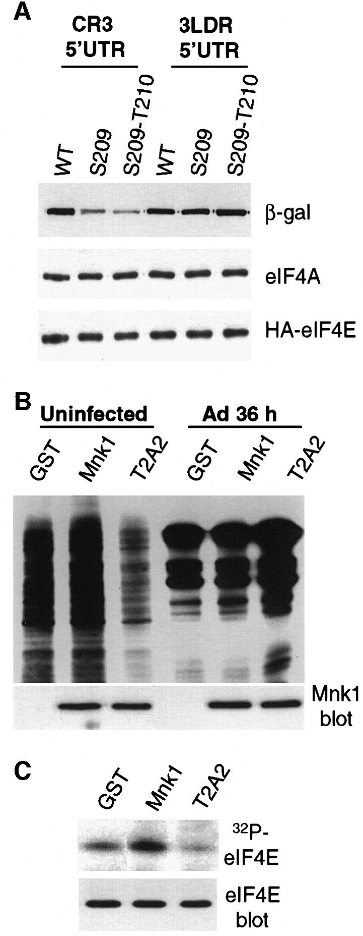
Fig. 4. Effect of eIF4E phosphorylation on translation of model Ad late and cellular mRNAs. (A) 293T cells were cotransfected with plasmids expressing β-galactosidase mRNAs containing either the Ad late tripartite leader 5′UTR (3LDR), or an eIF4F-dependent 5′UTR (CR3), and plasmids expressing HA-tagged wt eIF4E, Ser209→Ala eIF4E or Ser209→Ala/Thr210→Ala eIF4E. At 36 h post-transfection, equal amounts of cell lysates were resolved by SDS–12%PAGE and immunoblotted using specific antisera for β-galactosidase protein (β-gal), HA-eIF4E and eIF4A (for protein levels). (B) 293 cells were transfected with plasmids expressing GST, GST–Mnk1 or the GST–T2A2 Mnk1 mutant. At 18 h post-transfection, cells were infected with wtAd for 36 h, or were uninfected, then labeled with [35S]methionine, or (C) labeled with 32PO4. Samples were analyzed as in the legend to Figure 3. Mnk proteins were detected by immunoblot analysis with antisera to GST. eIF4E phosphorylation and abundance were examined as in the legend to Figure 3.