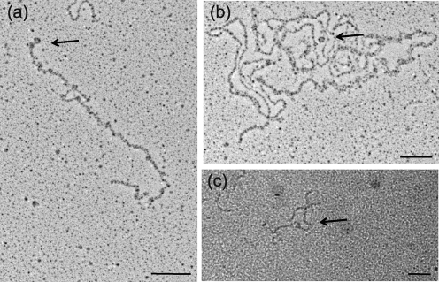
Fig. 8.
Ler bound to LEE5 DNA also forms circular structures. (a, b) Plasmid pKMTIR3 containing the LEE5 regulatory fragment (0.1 μg) with DNA from positions −303 to +172 was linearized by digestion with BamHI and then bound to purified Ler protein (0.5 μM) (a) or H-NS (1.0 μM) and Ler (0.5 μM) (b). After 15 min incubation at room temperature, samples were subjected to rotary shadowing and imaging by TEM as described in Methods. Bars, 100 nm. (c) The 475 bp LEE5 regulatory fragment (0.1 μg) containing DNA from positions −303 to +172 in relation to the transcriptional start site was bound to purified Ler protein at 0.5 μM, subjected to gold sputtering and viewed by TEM. Protein was not visible by gold sputtering. Arrows point to toroidal protein–DNA complexes. Bar, 50 nm.

