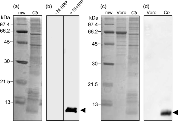
Fig. 1.
An ∼7.2 kDa component of C. burnetii binds Ni-HRP and is not present in host cells. (a) CBB-stained SDS-PAGE gel (12–15 % acrylamide; w/v) showing the protein profile of a whole-cell lysate from a mixed-cell population (10 days) of C. burnetii (Cb; 20 μg protein). Molecular mass markers (mw) are shown. (b) Corresponding FW blots of Cb, as in (a), showing an intense chemiluminescent band of ∼7.2 kDa (arrowhead) when probed with Ni-HRP (+ Ni-HRP; exposure time 2 min), but which is absent without Ni-HRP (−Ni-HRP; exposure time 5 min). (c) CBB-stained SDS-PAGE gel (12–15 % acrylamide; w/v) showing Cb and mw as above plus the protein profile of uninfected Vero host cells (Vero; 20 μg protein). (d) FW blot corresponding to Vero and Cb, as in (c), showing that the Ni-HRP binding component is specific to C. burnetii and is not detected in Vero cells.

