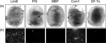
Fig. 8.
Immunoelectron and immunofluorescence microscopy showing the surface location of LimB in Coxiella. (a) Typical immunoelectron micrographs showing uniform protein A–gold binding to the surface of intact C. burnetii treated with anti-LimB or anti-Com1 (positive control for a surface-exposed protein) antibodies. Protein A–gold binding was rarely observed when negative controls were employed, including PIS, anti-MBP or anti-EF-Ts antibodies. (b) Typical immunofluorescence micrographs showing intense fluorescence at the surface of paraformaldehyde-fixed C. burnetii treated with antibodies against LimB or Com1 versus relatively minor, sporadic fluorescence when negative controls, as in (a), were employed.

