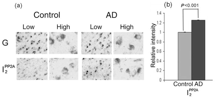
Figure 1.
Expression of I2PP2A mRNA in Alzheimer disease (AD) and control brain.27 (a) The I2PP2A signal was significantly elevated in AD brain (temporal cortex) compared with control brain (P < 0.001), whereas the GAPDH signal (G) was similar between the two. Differences between AD and control brains were analyzed statistically by Student’s t-test. High, high magnification (original magnification ×630); Low, low magnification (original magnification ×200). (b) The I2PP2A signals from five AD and five control cases were quantified and normalized against the GAPDH signal. Data show the mean ± SEM. Modified and reproduced with the permission from the American Society for Investigative Pathology from Tanimukai et al.27

