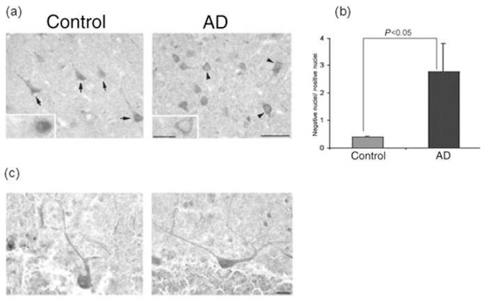
Figure 2.

Subcellular localization of I2PP2A in Alzheimer disease (AD) and control brains.27 (a) I2PP2A was predominantly expressed in the nucleus (arrows) of neurons in the temporal cortex from control brain, but was translocated from the nucleus to cytosol (arrowheads) in AD brain. (b) Ratio (mean ± SEM) of neurons with immunonegative to immunopositive nuclei. In AD brains, the number of neurons in the temporal cortex showing the translocation of I2PP2A from the nucleus to a cytoplasmic localization increased markedly (P < 0.05). Differences between AD and control cases were analyzed statistically by Student’s t-test. (c) In contrast, the subcellular localization of I2PP2A in the cerebellum was restricted to the nucleus in both AD and control brain. Modified and reproduced with the permission from the American Society for Investigative Pathology from Tanimukai et al.27
