
Figure 3.
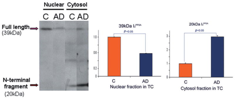
Cleavage and distribution of I2PP2A in nuclear and cytosolic fractions of the temporal cortex (TC) in Alzheimer disease (AD) and control (C) brains.27 Levels of I2PP2A in the nuclear fraction were decreased in AD compared with control brain. In contrast, the 39 kDa I2PP2A in the cytosolic fraction was decreased in AD brain, but the approximately 20 kDa fragment of I2PP2A was significantly increased in AD compared with control brain (*P < 0.05). Differences between AD and control brains were analyzed statistically by Student’s t-test. Modified and reproduced with the permission from the American Society for Investigative Pathology from Tanimukai et al.27