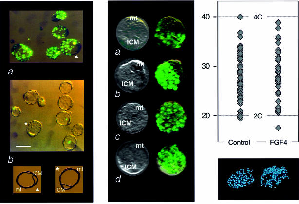
Fig. 4. Inhibition by FGF4 of the phagocytic activity and activation of DNA synthesis in mural trophectoderm cells. (A) Inhibition of phagocytosis. (a) Internalization of 1–2 µm fluorescent latex beads by 3.5 d.p.c. blastocysts (same experiment as in Figure 3A); (b) part of the same batch of embryos was maintained overnight in the presence of FGF4 (25 ng/ml; see Materials and methods). Tracing of one embryo in each series, indicated by arrowhead and star, respectively. Bar, 50 µm. (B) Activation of DNA synthesis in mural trophectoderm cells. 3.5 d.p.c. embryos were maintained overnight under the same conditions as in (A), either with (c and d) or without (a and b) the addition of 25 ng/ml FGF4. BrdU was then added to the medium for periods of either 2 (a and c) or 4 h (b and d) and incorporation was monitored by in situ immunodetection (mt, mural trophectoderm). (C) DNA content of blastocyst nuclei. Blastocysts were collected, maintained for 24 h in culture medium with (FGF4) and without (control) addition of 25 ng/ml FGF4. They were fixed and stained with Hoechst 33258 and nuclear DNA content was determined by fluorescence intensity reading performed on digitized images of individual nuclei, as previously described (Rassoulzadegan et al., 1998). Calibration of the diploid DNA content value was deduced from parallel measurements on spleen B lymphocytes (not shown). Top, values recorded for four blastocysts in the control and four in the FGF4 series. Bottom, fluorescence microscopy of one stained embryo in each series.

An official website of the United States government
Here's how you know
Official websites use .gov
A
.gov website belongs to an official
government organization in the United States.
Secure .gov websites use HTTPS
A lock (
) or https:// means you've safely
connected to the .gov website. Share sensitive
information only on official, secure websites.
