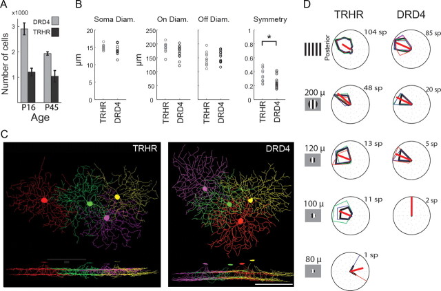
Figure 4.
Both TRHR- and DRD4-RGCs exhibit dendritic overlap which may compensate for their physiological properties. A, Cell counts of TRHR- and DRD4-RGCs from immunostained whole-mount retinas at P16 and P45. n = 5 for P16 DRD4 and n = 4 otherwise. B, Morphological properties of TRHR- and DRD4-RGCs revealed by Neurolucida reconstructions of filled cells. The parameters we measured corresponded to those previously reported to distinguish between mouse bistratified cells, and include soma size, dendritic diameters in On and Off layers, and symmetry assessment. All three diameters were not found as different (Kolmogorov–Smirnov test, p > 0.5, p > 0.14, and p > 0.74 for soma, On, and Off trees), while symmetry differed significantly (Kolmogorov–Smirnov test, p < 0.05). C, Neurolucida reconstructions of four neighboring TRHR-RGCs (right) and four neighboring DRD4-RGCs (left) that were sequentially filled. Bottom, XZ section of the reconstructed cells. Scale bar, 100 μm. D, Examples of directional tuning of TRHR-RGCs (middle column) and DRD4-RGCs (right column) in response to drifting gratings over a limited circular area. Left column illustrates the visual stimuli with the different diameters of gratings. The minimal area required for detection of motion was ∼100 μm and ∼125 μm for TRHR- and DRD4-DSGCs, respectively.

