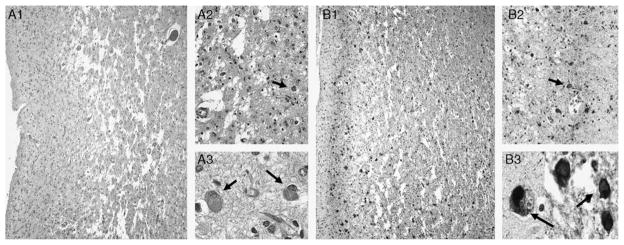
FIGURE 3.
Histologic sections. A, H&E staining. A1, Frontal cortex demonstrating spongiosis of the deep cortical layers (40 ×). A2, Highlights marked gliosis, neuron loss and Pick bodies (arrow) (400 ×). A3, Details of Pick bodies (arrows) (1000 ×). B1 and B2, τ-immunostaining of corresponding sections of frontal cortex showing abundant τ-immunoreactive residual neurons and Pick bodies (arrow) (40 × and 400 × , respectively). B3, τ-immunoreactive Pick bodies (arrows) (1000 ×). H&E indicates hematoxylin and eosin.

