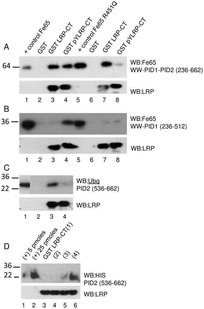
Figure 5.
LRP-CT binding to (A) Fe65 WW-PID1-PID2 236-662, (B) Fe65 WW-PID1 236-512, or (C) Fe65 PID2 536-662. GST-LRP-CT or GST-LRP-CT (pY4507) was immobilized on glutathione sepharose and 650 nM was incubated with 180–400 nM of each Fe65 construct (lanes 2–4) or Fe65(R451Q) construct (lanes 6–8) for 1 hour at 4°C. GST-coupled sepharose was used as a negative control. Bound Fe65 or Fe65(R451Q) was visualized by western blotting with anti-Fe65 antibody (Millipore, 3H6) or anti-Ubiqitin antibody (Invitrogen 13-1600). Each Fe65 variant (0.05 pmoles) was analyzed as a positive control (lane 1, 5). Equal LRP-CT loading is shown by western blotting with anti-LRP antibody (11H4). (D) LRP-CT binding to Fe65 PID2 536-662. GST-LRP-CT was immobilized on glutathione sepharose and 650 nM was incubated with 3 μM (1), 6 μM (2), 12 μM (3), or 24 μM (4) PID2 for 1 hour at 4°C. Bound Fe65 was visualized by western blotting with anti-HIS antibody (Quiagen, Penta-His Ab). Fe65 PID2 (5 and 25 pmol) is shown as a positive control. Equal LRP-CT loading is shown by western blotting with anti-LRP antibody (11H4).

