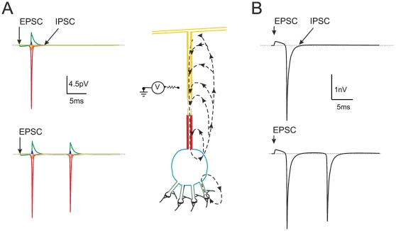
Figure 3. Extracellular field potential generated by a single granule cell.
(A) Schematic representation of a granule cell according to the model of Diwakar et al. [27]. The granule cell generates synaptic responses in the dendritic endings and action potentials in the axon hillock. This forms two current sinks, with the axon hillock giving by far the major contribution. The broken arrows depict the current flow, colors indicate the major neuronal comportments. The Na+ channels are concentrated in axon hillock, as indicated by immunohistochemistry [43], the excitatory and inhibitory synaptic channels are located in the terminal dendritic compartments. The circuit schematics on the left shows the flow of transmembrane current over the extracellular resistance. The extracellular potentials generated by different compartments of the granule cell model are shown to the left (same colors as in the neuron compartments). Notice that the extracellular potential is the largest in correspondence of the hillock, where Na+ channels have the highest density. (B) Extracellular field potential generated by a single granule cell “seen” from an electrode covering soma, dendrites and axon hillock (corresponding to a granular layer sink). Both in A and B, the neuron responds to the synchronous activation of all four mossy fibers (and all four inhibitory synapses, when active). Both in A and B, the neuron generates a single spike when synaptic inhibition is active, while it generates a doublet when synaptic inhibition is turned off.

