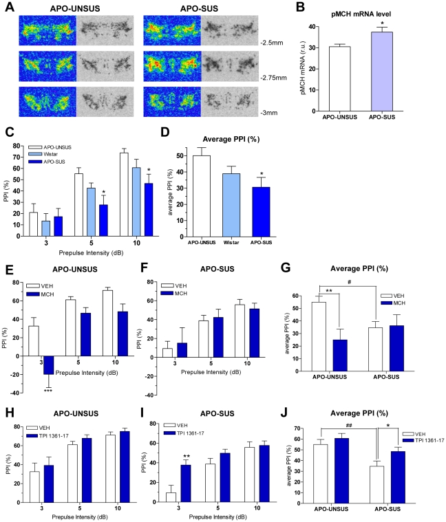
Figure 3. MCH effects on PPI in APO-UNSUS and APO-SUS rats.
A. Autoradiographic images illustrating the pMCH expression patterns in hypothalamic areas of APO-UNSUS and APO-SUS rats approximately at −2.5, −2.75 and −3 mm from bregma. B. pMCH mRNA levels in the lateral hypothalamus (LH) at −2.75 from bregma of APO-UNSUS and APO-SUS rats (*p<0.05 vs. APO-UNSUS, t-test; n = 5). C. PPI levels of naïve APO-UNSUS, wild type Wistar and APO-SUS rats (*p<0.05 vs. APO-UNSUS, two-way ANOVA with bonferroni test; n = 15). Values represent mean % PPI ± SEM. D. Average PPI level of naïve APO-UNSUS, wild type Wistar and APO-SUS rats (*p<0.05 vs. APO-UNSUS, one-way ANOVA with bonferroni test; n = 15). Values represent average of % PPI upon three prepulse intensities ± SEM. E. Effect of MCH (10 nmole) on PPI in APO-UNSUS rats (***p<0.001 vs. VEH, two-way ANOVA with Bonferroni test; n = 10–13). F. Effect of MCH (10 nmole) on PPI in APO-SUS rats (n = 10–13). G. Average of PPI values after VEH or MCH (10 nmole) injections in APO-UNSUS and APO-SUS rats (**p<0.01, # p<0.05 vs. VEH-treated APO-UNSUS rats, two-way ANOVA with Bonferroni test; n = 10–13). H. Effect of TPI 1361-17 (10 nmole) on PPI in APO-UNSUS rats (n = 12–13). I. Effect of TPI 1361-17 (10 nmole) on PPI in APO-SUS rats (**p<0.01 vs. VEH, two-way ANOVA with Bonferroni test; n = 13). J. Average of PPI values after VEH or TPI 1361-17 (10 nmole) injections in APO-UNSUS and APO-SUS rats (## p<0.01 vs. VEH-treated APO-UNSUS, *p<0.05 vs. VEH-treated APO-SUS, two-way ANOVA with Bonferroni test; n = 12–13). Data (E,F,H,I) are expressed as mean % PPI ± SEM. Data (G,J) represent average of % PPI elicited by three prepulse intensities ± SEM.

