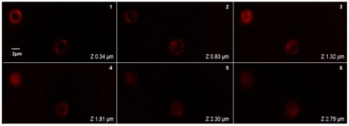
Figure 3. Localization of AF-SWCNTs in side erythrocytes by confocal microscopy.
Fluorescenated AF-SWCNTs were prepared as described in Materials and Methods. Erythrocytes were incubated in RPMI +1%FBS with 50 µg/ml fluorescenated AF-SWCNTs for 1 h and unbound/loosely attached particles were removed by three extensive washings with PBS. After that erythrocyte suspension was taken for confocal microscopy. Above figure shows fluorescence images of erythrocytes incubated with alexa fluor 633 hydrazide tagged AF-SWCNTs as z-sections. (Magnification 100X).

