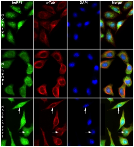
Figure 4. Subcellular localization of human eRF1 fused with short or long N-terminal sequence of Ilf3/NF90.
Plasmids pCMV-heRF1-Ilf3/NF90 short N-terminal sequence (N-heRF1, mid panels) and pCMV-heRF1-Ilf3/NF90 long N-terminal sequence (NoLS-heRF1, lower panels) were transfected into HeLa cells. After 24 hours, untransfected (Control, upper panels) or transfected cells were co-stained with anti-heRF1 antibody (heRF1), anti-α-tubulin antibody (α-Tub) and DAPI. Endogenous heRF1 or heRF1 recombinant fusion proteins appear in green, α-tubulin in red and DAPI in blue. Arrows point to intranuclear foci corresponding to nucleoli.

