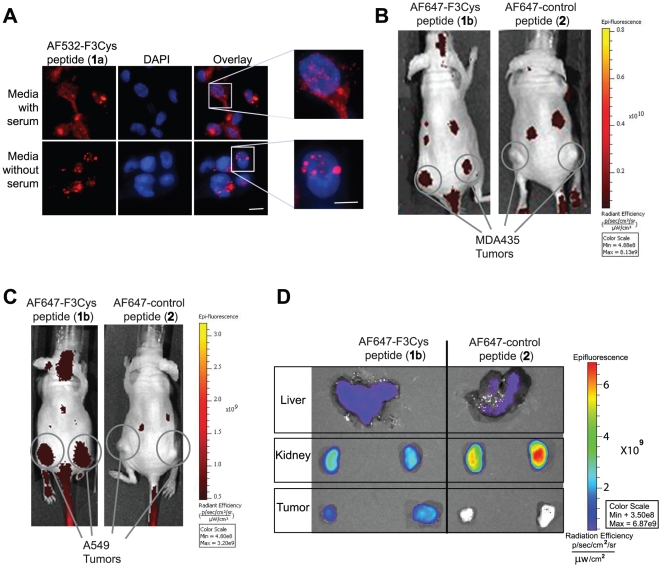
Figure 2. In vitro and in vivo optical imaging using Fluorescent-labeled F3Cys peptides.
MDA-MB-435 cells, in optically clear bottom dishes, cultured in either serum free or serum containing media were stained with AF532-F3Cys, counterstained with DAPI and monitored under a fluorescent microscope (A). Mice bearing MDA-MB-435 (B) or A549 (C) xenografts were injected i.v. via the tail vein with either AF647-F3Cys (1b) or AF647-Control peptide (2). Fluorescence images were acquired over time and a representative image obtained at 2 h is shown (B and C). Ex vivo fluorescence imaging of tumor, kidney and liver harvested 2 h after AF647-F3Cys peptide injection in animals bearing A549 tumor xenograft (D).

