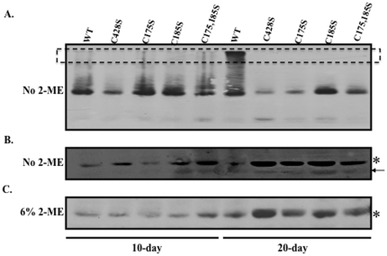
Figure 4. Nonreducing Western blot analyses of 10 and 20-day wild-type and mutant viruses.
Representative Western blot images of equal volumes of 10 and 20-day wild-type and mutant viruses run under nonreducing (A–B) and reducing (C) conditions. Monomeric L1 (lower band in A) under nonreducing conditions is shown overexposed in (B). Monoclonal H16.J4 was used to detect HPV16 L1. Multimeric L1 species are indicated by a box. An arrow points to a faster migrating L1 species which has been previously shown to be present in greater concentration within purified virions [23].

