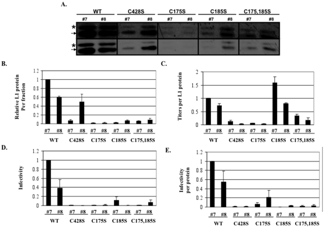
Figure 7. Analysis of infectious Optiprep fractions.
(A) Western blot analysis of L1 in highly infectious Optiprep fractions #7 and #8 from Optiprep-fractionated 20-day wild-type (WT), C428S, C175S, C185S, and C175,185S viruses. The two rows are replicates with the top being overexposed. Higher and lower molecular weight species of L1 are indicated. (B) Densitometric analysis of the faster migrating L1 species (i.e. 55 kD) in three independent Western blots as shown in A. WT fraction #7 was set to 1.0. (C) Titers per fraction, normalized to the relative amount of L1 protein within fractions. WT fraction #7 was set to 1.0. (D) Infectivity of 20-day wild-type (WT), C428S, C175S, C185S, C175,185S virions within fractions #7 and #8. WT is set to 1.0. (E) Infectivity of 20-day wild-type (WT), C428S, C175S, C185S, C175,185S virions within fractions #7 and #8. Relative L1 protein content per fraction were used to normalize relative infectivity data within fractions. WT is set to 1.0.The results are means and standard errors for at least three independent experiments. The arrow points to the L1 band with the same motility as seen with L1 from PsV [23]. The asterisk marks the slow mobility L1 band seen only in native HPV16 virion [23].

