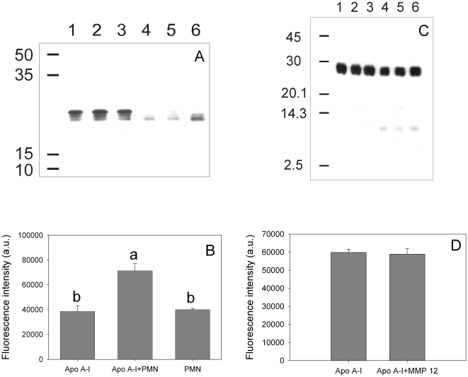
Figure 6. Effect of TPA-activated neutrophils and MMP-12 on apoA-I processing and amyloidogenicity.
ApoA-I (0.2 mg/mL) was incubated in the presence of TPA-stimulated neutrophils at pH 7.4 at 37°C for 1 h. (A) Western blotting of aliquots of the supernatant developed with rabbit polyclonal anti-apoA-I. Lanes 1 to 3 show samples incubated in the absence of neutrophils, while lanes 4 to 6 represent the same amount of protein applied to each lane after neutrophil treatment. (B) ThT binding to apoA-I previously incubated (24 h at 37°C) in the absence (labeled as ApoA-I) or in the presence of TPA-stimulated neutrophils (ApoA-I + PMN). As an additional control, ThT fluorescence associated to neutrophils incubated under the same conditions without apoA-I is also shown (bar labeled PMN). In a different experiment apoA-I (0.2 mg/mL) was incubated in the presence of MMP-12. (C) Western blotting was developed as in (A). Numbers on the left side of the figures (A) and (C) correspond to Molecular Weight Markers (GE Healthcare, Uppsala, Sweden). (D) After 3 h at 37°C, MMP-12 was inhibited and apoA-I incubated as in (B) to determine ThT binding. Bars correspond to means ±SE. Bars denoted by different letters differ significantly at p<0.05.

