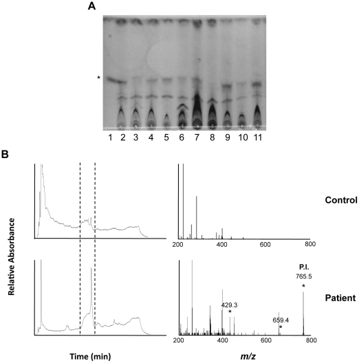
Figure 4. Structurally intact mycolactone is detected in serum samples of BU patients.
(A) Example of the fluorescence signals given by serum samples (lanes 2–9), following lipid extraction and analysis by TLC-Fluo. Controls include 1 µg pure mycolactone (lane 1), lipids extracted from a negative serum (lane 10), and then spiked with 1 µg mycolactone (lane 11). The band corresponding to mycolactone is designated by an asterisk. (B) Representative HPLC elution profiles of lipids extracted from serum samples are shown for one healthy control out of 5, and one BU patient among the 4 positive ones. The corresponding MS/MS spectra show the presence of the parent and product ions of mycolactone in this positive sample. Similar results were obtained in a second one.

