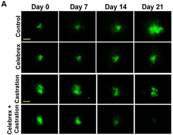
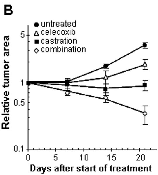
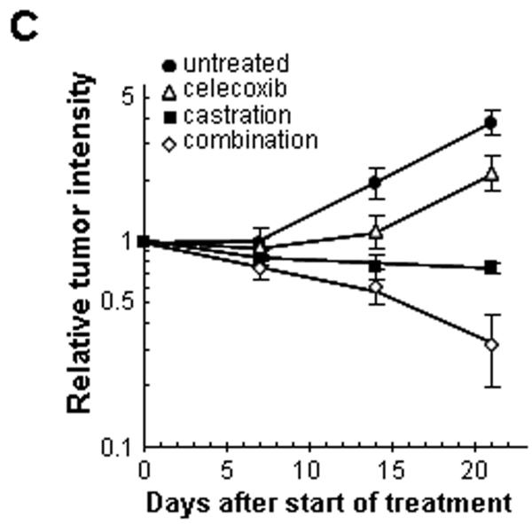
Figure 3. Effect of celecoxib and/or surgical castration on prostate tumor growth in vivo.
TRAMP-C2-GFP cell spheroids were co-implanted with prostate tissue and allowed to vascularize. When there was proper blood flow within the growing tumors, the mice were surgically castrated (Day 0) and Celecoxib treatment (15 mg/kg/administration) was started by oral administration twice daily. Tumors were imaged by intravital microscopy once a week. Panel A: a representative collage of tumor growth in the four treatment groups. Bar ~ 500μm. Panel B-C: graphic representation of relative tumor areas (B) and relative tumor intensities (C) calculated from intravital microscopy data (log scale).


