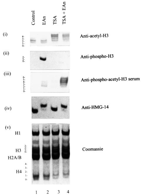
Fig. 4. Generation of antibodies against doubly modified phosphoacetyl histone H3. Acid-soluble nuclear proteins were extracted from quiescent C3H 10T1/2 cells (lane 1) or cells stimulated with 50 ng/ml EGF and 10 µg/ml anisomycin for 1 h (EAn, lane 2), pretreated with 500 ng/ml TSA for 4 h (lane 3) or pretreated with 500 ng/ml TSA for 4 h and then stimulated with EGF/anisomycin for the last hour (lane 4) and electrophoresed on 15% acid–urea gels. Proteins were transferred to PVDF membrane and analysed by western blotting using anti-acetyl-H3 antibodies (panel i), anti-phospho-H3 antibodies (panel ii), anti-phosphoacetyl-H3 antibodies (panel iii) or anti-HMG-14 antibodies (panel iv). Coomassie-stained gel is shown in panel v. The positions of the modified forms of histone H3 are numbered corresponding to the number of post-translational modifications visualized by Coomassie staining of gels or Ponceau S staining of PVDF membrane.

An official website of the United States government
Here's how you know
Official websites use .gov
A
.gov website belongs to an official
government organization in the United States.
Secure .gov websites use HTTPS
A lock (
) or https:// means you've safely
connected to the .gov website. Share sensitive
information only on official, secure websites.
