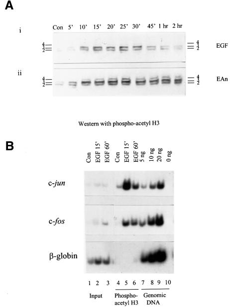
Fig. 8. Phosphoacetyl-H3 is associated with c-jun and c-fos chromatin upon physiological stimulation. (A) Quiescent control (Con) C3H 10T1/2 cells were stimulated with EGF (50 ng/ml) alone or with EGF (50 ng/ml) and anisomycin (10 µg/ml) for the times indicated. Acid-soluble nuclear proteins were resolved on 15% acid–urea gels, transferred to PVDF membrane and probed with the anti-phosphoacetyl-H3 antibody. (B) Formaldehyde cross-linked chromatin was prepared from quiescent (Con) and EGF (50 ng/ml)-stimulated mouse C3H 10T1/2 cells as described in Materials and methods, and immunoprecipitated with anti-phosphoacetyl-H3 antibodies (lanes 4–6). The recovered DNAs from the antibody-bound fractions, the total input DNA from released chromatin used for the ChIP assays (Input, lanes 1–3) and genomic DNA (lanes 7–9; lane 10 lacks template) were analysed for the presence of c-jun (upper panel), c-fos (middle panel) and β-globin (lower panel) sequences by PCR as described in the legend to Figure 7.

An official website of the United States government
Here's how you know
Official websites use .gov
A
.gov website belongs to an official
government organization in the United States.
Secure .gov websites use HTTPS
A lock (
) or https:// means you've safely
connected to the .gov website. Share sensitive
information only on official, secure websites.
