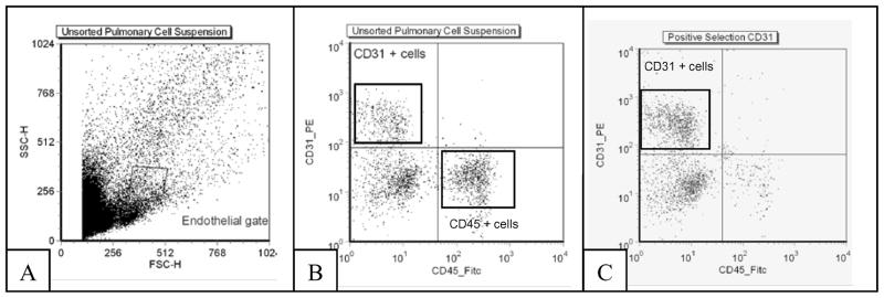
Figure 2. Magnetic Isolation of Pulmonary Endothelial Cells.
Whole lung tissue from mice (n = 5 mice per surgical group) was pooled and homogenized into a single cell suspension and pulmonary endothelial cells isolated using a microbead sorting system. (A) Total cells prior to sorting, without fluorescent antibody label. (B) Cells stained with CD45-FITC and CD31-PE (C) Pure population of CD31(+), CD45(−) cells after a negative sort for CD 45 and a second, positive selection for CD31.

