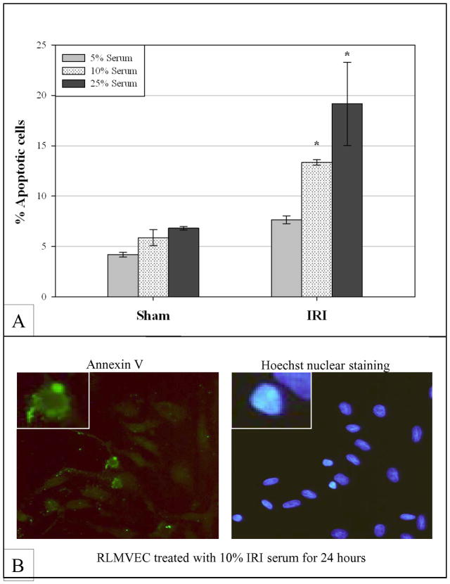
Figure 5. AKI Induces Apoptosis in Vascular Endothelial Cells.
Rat Lung Microvascular Endothelial Cells (RLMVECs) were cultured in the presence of serum from animals that underwent either bilateral IRI or sham surgery. Serum was isolated 24 hours after injury; cells were then cultured in either 5%, 10% or 25% serum for 24 hours. After serum exposure, cells were stained with a FITC-conjugated antibody against Annexin V as well as PI and then evaluated by FACS. Cells exposed to IRI serum showed increased evidence of apoptosis in a dose-dependent manner (A) compared to cells exposed to sham serum. There was a statistically significant difference in apoptosis between IRI and Sham in the 10% and 25% serum groups. Notably there was no significant difference in apoptosis amongst the sham groups. (B) An RLMVEC stained with Annexin V and Hoechst for nuclear visualization reveals membrane localization of Annexin V as well as nuclear changes consistent with apoptosis.

