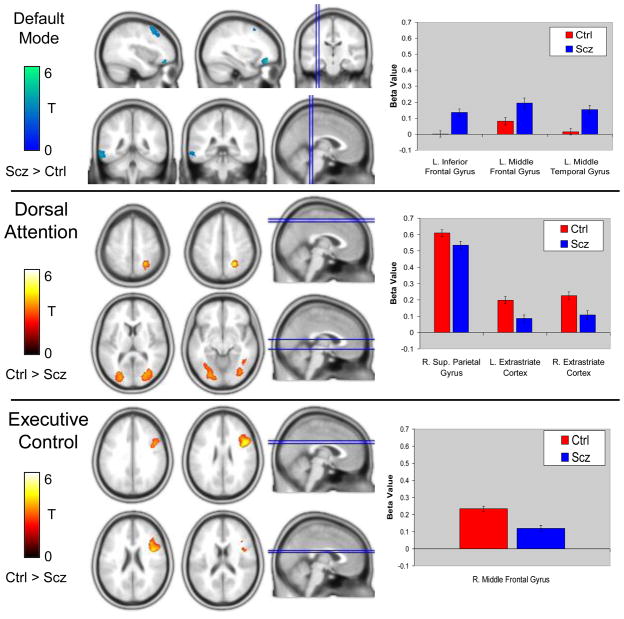
Figure 2.
Resting-state functional connectivity differences between schizophrenia patients and healthy control subjects in the default mode, dorsal attention, and executive control networks. Compared to control subjects, patients demonstrated greater connectivity between the PCC seed ROI and the left inferior frontal gyrus, left middle frontal gyrus, and left middle temporal gyrus (Top Panel). In contrast, patients demonstrated reduced connectivity between the dorsal attention IPS/SPL seed ROI and right superior parietal gyrus and bilateral extrastriate visual areas (Middle Panel). Similarly, connectivity between the executive control dlPFC seed ROI and right middle frontal gyrus was reduced in schizophrenia (Bottom Panel). Abbreviations: Ctrl=Control group; L=Left; R=Right; Scz=Schizophrenia group.

